
Applications of optical tweezers

Intracellular rheology
for cell elasticity and
viscosity measurements

Characterize the mechanical state of cells measuring their stiffness, elasticity and viscosity and obtain the complex shear modulus of the cellular medium using the built-in active TimSom micro rheology routine of our SENSOCELL optical tweezers.


Intracellular active rheology

Probe the strain response of intracellular medium as a function of frequency with our automatized TimSom  Active Rheology routine and obtain the storage (G′, elastic contribution) and loss (G″, viscous contribution) moduli of cell cytoplasm.

Cytoplasm and nucleus rheology inside a Zebra fish progenitor cell

In this example, the SENSOCELL optical tweezers were used to measure the viscoelastic properties of Zebra fish stem cells ex vivo. The researchers used the TimSOM active rheology routine to obtain the complex shear modulus G at different locations of the cell: at the cell’s cytoplasm, the nuclear interface and inside the cell nucleus.


Experimental:

Quantify cell stiffness, viscosity and elasticity

Our SENSOCELL optical tweezers allow measuring the rheological properties of cells inside tissues, embryos and living organisms such as Zebra fish or C. elegans systems.

Perform active rheology assays of cells trapping either internalized microspheres or endogenous organelles such as vesicles or lipid droplets as probes.

The Sensocell optical tweezers workflow allows applying oscillations to the trapped probes with frequencies ranging from 0.01 Hz up to 6 kHz and automatically computes the complex shear modulus G of the sample at the selected frequencies.

Selected publications:

Nature physics

Nat. Phys. 17, 1270–1276 (2021)

RHurst, S., Vos, B.E., Brandt, M. et al.

Intracellular softening and increased viscoelastic fluidity during division.

Nat. Mater. 22, 1409–1420 (2023)

Kechagia, Z., Sáez, P., Gómez-González, M. et al.

The laminin–keratin link shields the nucleus from mechanical deformation and signalling.

Biorxive 2023

Frederic Català-Castro, Santiago Ortiz-Vásquez, Carmen Martínez-Fernández et al.

Active microrheology with a single, time-shared laser trap.
Application Note:

Micro-rheology of soft biological samples using SENSOCELL optical tweezers.

Related applications:

Sensocell case studies

In vivo cytoplasm & nucleus rheology inside a living c. elegans organism

Quantifcation of the complex shear modulus of Caenorhabditis elegans changes during age using TimSOM Active Rheology with Sensocell optical tweezers. Using mutants known to display premature ageing phenotypes in C. elegans, M. Krieg’s group revealed how mutations causing nuclear envelopathies soften the cytosol of intestinal cells during organismal age. The data shows the frequency dependent shear modulus for two different ages of wildtype and age-matched lem-2 mutants and how the rheological properties of the cellular cytoplasm evolve during the first eight days of age. For these measurements, endogenous lipid droplets were used as mechanical stress probes. The viscosity of the cytoplasm was extracted from the fit of the fractional Kelvin-Voigt model to the rheological spectrum data. See more details in Biorxive 2023. Courtesy of M. Krieg’s lab (ICFO).

Related applications:

Intracellular active rheology inside a yeast cell

Full video of a TimSom active micro-rheology experiment carried out inside a living yeast cell with SENSOCELL optical tweezers. The cell’s vacuole was optically trapped and used as a probe to obtain the energy storage G’ and energy loss G” moduli of the cellular medium in the frequency range of 0.5 Hz – 1kHz. The integrated data analysis software allows fitting the data with several biophysical models like Maxwell or Kelvin-Voigt.

See other video examples:
CONCEPTS / ADVANTAGES

Key concepts & Advantages

Key concepts

  • Rheology: a field of material science that studies the flow behavior of viscoelastic fluids. In cell biophysics, it helps explain how the plasma membrane responds to tension and how stress is distributed throughout the cytoplasm during the cell cycle.


  • Shear Modulus (G): describes a material’s response to shear stress. It is related to the Young’s modulus by the equation G=E/2(1+v) where v is the Poisson ratio


  • Strain (ϵ): a dimensionless parameter representing the normalized deformation that an object undergoes when subjected to stress.


  • Stress (σ): the amount of force per unit area exerted on an object, measured in units of pressure (Pa).


  • Viscoelasticity: the property of materials to exhibit both viscous and elastic characteristics simultaneously, meaning they partially withstand stress.

Advantages

  • Precision: SENSOCELL’s high-resolution measurements allow for detailed characterization of the complex shear modulus of cellular medium and its viscoelastic properties.


  • Non-destructive: these experiments can be minimally invasive, preserving cell viability and function.


  • Automatized routine: the TimSom active rheology routine integrated in the SENSOCELL system offers an automatized workflow for rheological assays including fitting analysis of the computed G modulus data.


  • Wide spectral range: active rheology tests deliver dynamic information spanning over five decades of frequencies.

Conclusions

The TimSom active rheology routine integrated in the Sensocell optical tweezers is a unique tool for measuring the rheological properties of the intracellular medium, delivering key information of its viscoelastic properties over a wide spectral range.

Download Full Active Micro-Rheology Application Note:

Micro-rheology of soft biological samples using optical tweezers.
(Clic image to download)
Video 1

Confocal video showing a DVA neuron Ca2+ ion channel activity in a dynamic optical trapping assay. The pulling rate applied to the membrane tether is progressively increased. Scale bar = 5µm. Acquired at 10Hz.

Fig. 1

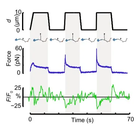
Time dependence of trap position (top), force signal (middle) and Ca2+ fluorescence signal (bottom) recorded during a membrane tether pulling experiment. The force and Ca2+ ion channel activity signals exhibit negative correlation.

Fig. 2

Force and displacement data for two beads stimulated at frequencies of 4 Hz and 0.25 Hz. The progressive reduction of the beads displacement is in concomitancy with an increase of the applied force and loading rate.

Fig. 1

Schematic representation of the optical tweezers experiment. A fibronectin-coated bead is trapped, brought into contact with the cell membrane and stimulated with oscillations at different frequencies.

Fig.2

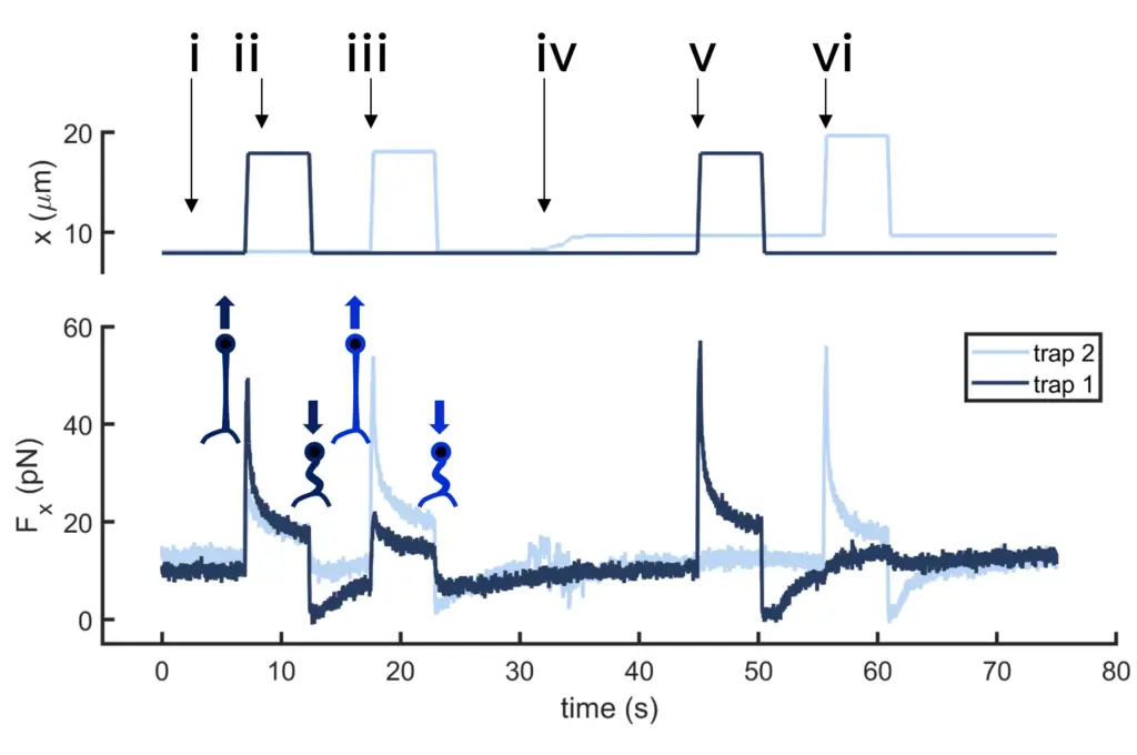
Time dependence of trap 1 and trap 2 position (top) and force (bottom) data along the different steps of the described dual tether pulling experiment.

Fig.1

Confocal video of the described dual tether pulling assay performed on a neuron axon. Courtesy of M. Krieg lab (ICFO).

Fig. 1

Time dependence of trap position (top), force signal (middle) and Ca2+ fluorescence signal (bottom) recorded during a membrane tether pulling experiment. The force and Ca2+ ion channel activity signals exhibit negative correlation.