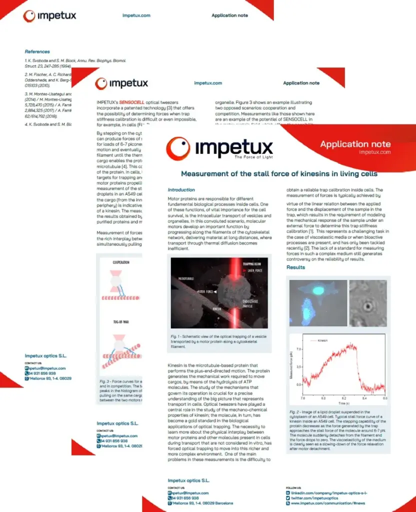
Optical tweezers are a powerful tool for studying the activity of motor proteins like kinesin, myosin, or dynein and characterize the mechanical properties of microfilaments and microtubules.
Optical tweezers are a key tool for understanding how motor proteins like kinesin, dynein or myosin contribute to intracellular transport and organization. Also, the study of the mechanical properties of cytoskeletal filaments such as actin and microtubules is essential for understanding their roles in cellular structure and dynamics. These applications have a strong impact in disease research since studying the effects of mutations or drugs on filament mechanics and motor protein activity is relevant for diseases involving cytoskeletal dysfunctions.
SENSOCELL optical tweezers enable the study of single-molecule motor proteins in isolation, providing insights into their fundamental properties without the interference of bulk effects. Furthermore, thanks to its direct force sensor technology, Sensocell extends the study of motor protein activity from single-molecule assays to experiments in living cells.
A typical single-molecule motor protein experiment consists of different steps:
Typically, motor proteins are attached to a functionalized microscopic bead. The bead is then trapped by the optical tweezers.
The motor protein interacts with its substrate (microtubules for kinesin and dynein, actin filaments for myosin), which can be immobilized on a surface.
As the motor protein moves along the filament, the movement of the bead is tracked with high precision to study the motor’s stepping behavior and force generation.
Optical tweezers are also highly effective for studying the mechanical properties of cytoskeletal filaments such as actin and microtubules. Potential applications include measuring the filaments stiffness by stretching assays or doing rheological measurements to quantify the viscoelastic behavior of cytoskeletal filaments. Common experimental steps for such assays are:
Actin filaments or microtubules are polymerized and often labeled with fluorescent markers for visualization.
Microscopic beads are attached to the ends or specific points along the filaments. These beads are then trapped using the optical tweezers.
Controlled forces are applied using the tweezers, and the resulting displacements are measured with high precision to study the mechanical response of the filaments.

Reddy, B., Mattson, M., Wynne, C. et al.
Load-induced enhancement of Dynein force production by LIS1–NudE in vivo and in vitro.Microtubules motor proteins are responsible for different fundamental biological processes inside cells. One of these functions, of vital importance for the cell survival, is the intracellular transport of vesicles and organelles along microtubules. Kinesin is the microtubule-based motor protein that performs the plus-end-directed motion. The protein generates the mechanical work required to move cargos, by means of the hydrolysis of ATP molecules.
We can use lipid droplets as targets for trapping and analysis of the force of the motor proteins propelling them inside living cells. Fig.1 shows the measurement of the stall force of a kinesin motor protein transporting lipid droplets along a microtubule in a A549 cell.
Measurement of forces inside cells allows exploring the rich interplay between multiple molecular motors simultaneously pulling on the same vesicle/organelle. Here we show an example illustrating two opposed scenarios: cooperation and competition. This is an example of the potential of SENSOCELL optical tweezers in the motor proteins field, which offers the possibility of measuring forces in traditionally difficult or impossible experiments.
SENSOCELL optical tweezers provide a versatile and powerful method for studying motor protein activity and cytoskeletal filaments mechanics, providing precise and quantitative insights into their mechanical properties at the single-molecule level.
Confocal video showing a DVA neuron Ca2+ ion channel activity in a dynamic optical trapping assay. The pulling rate applied to the membrane tether is progressively increased. Scale bar = 5µm. Acquired at 10Hz.
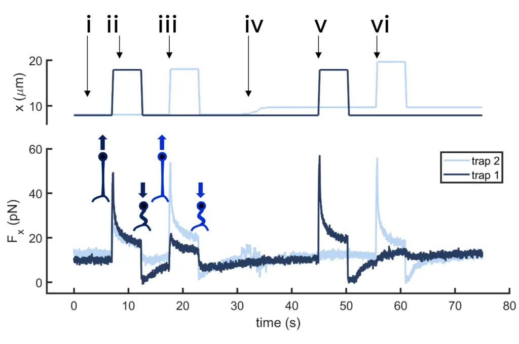
Time dependence of trap 1 and trap 2 position (top) and force (bottom) data along the different steps of the described dual tether pulling experiment.
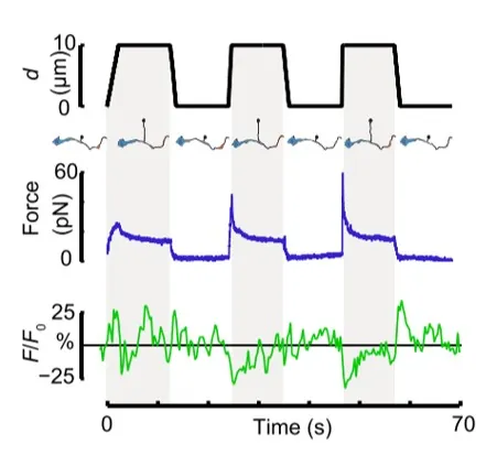
Time dependence of trap position (top), force signal (middle) and Ca2+ fluorescence signal (bottom) recorded during a membrane tether pulling experiment. The force and Ca2+ ion channel activity signals exhibit negative correlation.
Force and displacement data for two beads stimulated at frequencies of 4 Hz and 0.25 Hz. The progressive reduction of the beads displacement is in concomitancy with an increase of the applied force and loading rate.
Schematic representation of the optical tweezers experiment. A fibronectin-coated bead is trapped, brought into contact with the cell membrane and stimulated with oscillations at different frequencies.
Confocal video of the described dual tether pulling assay performed on a neuron axon. Courtesy of M. Krieg lab (ICFO).
Time dependence of trap position (top), force signal (middle) and Ca2+ fluorescence signal (bottom) recorded during a membrane tether pulling experiment. The force and Ca2+ ion channel activity signals exhibit negative correlation.