
SENSOCELL optical tweezers provide precise measurement of cell-cell interaction forces, facilitating a deeper insight into immune interactions, disease mechanisms, and cellular signalling.
Understanding cell-cell interaction forces is key for studying immune interactions, as these forces play a vital role in various physiological and pathological processes. Optical tweezers provide a powerful tool for trapping and manipulating cells, while direct force detectors based on light momentum analysis enable precise measurement of the forces exerted on the cells.
First, we use the optical tweezers to trap and hold both cells in place using one optical trap for each cell. Next, the optical tweezers apply controlled trap motion to bring the cells into contact. SENSOCELL optical tweezers allow for precise control over the distance and orientation between the interacting cells and its integrated direct force detector allows measuring the interaction forces as the cells touch and interact. After a predefined interaction time, we move the cells apart and continue to measure the forces as they detach, thereby enabling the measurement of cell-cell adhesion force. Finally, the collected force and position data are used to generate force-distance and force-time curves, which provide detailed insights into the interaction forces at various stages of contact.

Glass DG, McAlinden N, Millington OR, Wright AJ.
A minimally invasive optical trapping system to understand cellular interactions at onset of an immune response.In collaboration with Dr. Carlos Barcia Lab at the Universitat Autònoma of Barcelona (UAB), we demonstrate how Sensocell optical tweezers enable precise control of a trapped neuroblastoma cancer cell and a T-cell using two optical traps. In this example, we move the T-cell towards the cancer cell using the manual “click and drag” mode, establishing cell-cell contact. Next, after 10 seconds, the tweezers pull the T-cell back until the adhesion bond breaks. Throughout the process, the system records force and position data.
The accompanying figure and video illustrate the force dependence over time for the trap holding the T-cell. When the applied force is sufficient to break the bond between the two cells, the measured force drops to zero. In this case, the measured cell-cell adhesion force was 21 pN, consistent with the expected adhesion force of a single T-cell receptor
Chimeric Antigen Receptor T-cells (CAR-T cells) are genetically engineered T-cells designed to target and destroy cancer cells. SENSOCELL optical tweezers enable studying the interaction forces between CAR-T cells and cancer cells with high precision controlling parameters such as the interaction time and orientation. In this way, we can measure the adhesion force when the CAR-T cell binds to the cancer cell, its binding probability and detect multiple binding and unbinding events.
This information can help understand the biophysical mechanisms of how CAR-T cells recognize, bind to, and kill cancer cells. Insights gained from these measurements can help predict the success of CAR-T cell therapies by understanding the strength and stability of the interactions between different CAR-T cells and cancer cells. This is a valuable knowledge for developing new drugs that can enhance the effectiveness of CAR-T cell therapies by modulating their interaction forces.
In this example in collaboration with Dr. Juan Manel Otero from the Hospital Clínic of Barcelona, we used SENSOCELL optical tweezers to quantify cell-cell interactions between CAR-T cells and Lymphoma cancer cells. The cell-cell interaction time was set to 15 seconds using automated trap motion routines. The obtained interaction force traces often display multiple unbinding events. For example, here we observe two distinct unbinding events (see Fig. 1).
Sensocell optical tweezers, with its direct force detectors based on light momentum analysis, provide a powerful and precise method for studying cell-cell interaction forces. Its distinctive technology allows measuring forces on cells and offers significant advantages in precision, sensitivity, and non-invasive measurement capabilities.
Confocal video of the described dual tether pulling assay performed on a neuron axon. Courtesy of M. Krieg lab (ICFO).
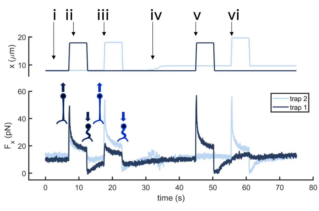
Time dependence of trap 1 and trap 2 position (top) and force (bottom) data along the different steps of the described dual tether pulling experiment.
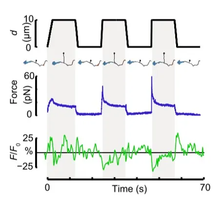
Time dependence of trap position (top), force signal (middle) and Ca2+ fluorescence signal (bottom) recorded during a membrane tether pulling experiment. The force and Ca2+ ion channel activity signals exhibit negative correlation.
Force and displacement data for two beads stimulated at frequencies of 4 Hz and 0.25 Hz. The progressive reduction of the beads displacement is in concomitancy with an increase of the applied force and loading rate.
Schematic representation of the optical tweezers experiment. A fibronectin-coated bead is trapped, brought into contact with the cell membrane and stimulated with oscillations at different frequencies.
Time dependence of trap position (top), force signal (middle) and Ca2+ fluorescence signal (bottom) recorded during a membrane tether pulling experiment. The force and Ca2+ ion channel activity signals exhibit negative correlation.
Confocal video showing a DVA neuron Ca2+ ion channel activity in a dynamic optical trapping assay. The pulling rate applied to the membrane tether is progressively increased. Scale bar = 5µm. Acquired at 10Hz.