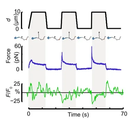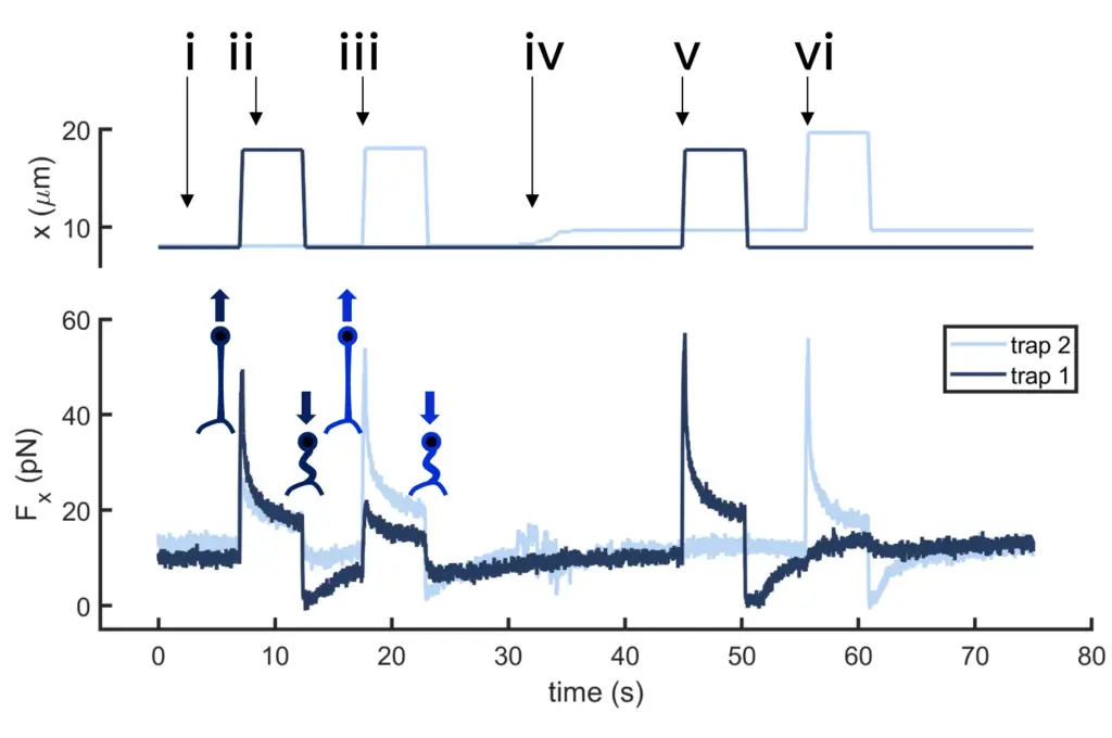
Cell motility and cell
motion force dynamics
Study cell motility of sperm cells, flagellated bacteria or other microswimmers and quantify the cell motion force dynamics governing their movement with SENSOCELL optical tweezers.
Cell motility and cell motion force quantification
SENSOCELL optical tweezers can track the movement of motile cells, measuring forces generated by the cell during active movement. Each cell can be individually analyzed for its mechanical properties and motility behavior.
How to perform cell motility studies with SENSOCELL
Explore the dynamics of cell motility using SENSOCELL optical tweezers and quantify cell motion forces using its calibration-free force spectroscopy module. Additionally, the built-in active microrheology routine in SENSOCELL allows measuring the medium viscosity and study its influence on your cell motility assays.
Trapping the target cells
Optical tweezers can trap and hold one or multiple target cells, using one optical trap per cell.
Real-time force detection
SENSOCELL’s force detector provides dynamic, real-time data of the forces applied on eah cell preventing them to escaping their respective optical trap.
Analyzing data
Forces measured in the XY plane help elucidate complex cell motility behaviors, such as oscillatory force dynamics. Additionally, the instrument provides valuable data in the frequency domain for thermodynamic studies.
Selected publications:
APS Journals

Phys. Rev. E (2021)
Corbyn Jones, Mauricio Gomez, Ryan M. Muoio, Alex Vidal, Anthony Mcknight, Nicholas D. Brubaker, Wylie W. Ahmed.
Stochastic force dynamics of the model microswimmer Chlamydomonas reinhardtii: Active forces and energetics.Sensocell case studies
Cell motion force dynamics of microswimmer Chlamydomonas reinhardtii
This application example is part of a work from Wylie Ahmed’s Lab (Toulouse University). In this work, the authors demonstrate direct measurement of cell motion forces generated by Chlamydomonas microswimmers and interpreted their motion dynamics using simulations of self-propelled particles.
For this purpose, the authors used a custom optical trapping instrument designed by Impetux to experimentally measure the stochastic force dynamics of a Chlamydomonas microswimmer. Force measurements were performed using our light momentum based force spectroscopy module. Analyzing force and active energy spectrum data of a swimming Chlamydomonas and following a thermodynamic approach, the authors quantified the non-equilibrium activity of the microswimmer.
The authors show that Chlamydomonas exhibit complex oscillatory force dynamics with magnitude of tens of pNs, and rotational dynamics of 1-2 Hz, with an an average power dissipation of approximately 5 fW.
Key concepts & Advantages
Key concepts
Cell Motion: The movement of cells from one location to another, which can be either active (due to the cell’s own mechanisms) or passive (due to external forces).
Cell Motility: The ability of cells to actively move and navigate their environment using energy-driven processes.
Microswimmer: A microscopic organism or synthetic particle capable of self-propulsion in a fluid environment, often studied to understand locomotion at small scales, such as bacteria, sperm cells, or engineered nano-robots.
Advantages
- DIrect Force measurements: SENSOCELL’s direct force sensor enables the measurement of forces applied to cells without requiring prior calibration.
- Sensitivity: SENSOCELL can measure very small forces in the picoNewton (pN) range up to several hundreds of pN, which is ideal for detecting motion forces of microswimmers.
- Cell viability: optical tweezers are minimally invasive, reducing potential damage to the cells and assuring cell viabiility throughout the experiment.
- Temporal resolution: the ability to measure forces in real-time at high temporal resolution provides dynamic insights into the processes governing cell motility.
Conclusions
The extensive trapping and force detection capabilities of SENSOCELL optical tweezers enable the simultaneous measurement of cell motion forces for multiple microswimmers, regardless of their shape and size, thus making it an ideal tool for cell motility assays.