PHASESTREAK CONFOCAL

Fast deep confocal imaging
Beyond mechanical constraints

PhaseStreak is designed to adapt to different samples and imaging requirements. It combines fast acquisition, flexible control over illumination and strong optical sectioning in a robust platform designed for minimal routine maintenance.

01 / KEY FEATURES

Rethinking confocal imaging for speed and flexibility

PhaseStreak is designed to adapt to different samples and imaging requirements. It combines fast acquisition, flexible control over illumination and strong optical sectioning in a robust platform designed for minimal routine maintenance.

SOLID-STATE ARCHITECTURE

No mechanical scanners or physical pinholes.

230 FPS

Camera-limited confocal and widefield acquisition.

3 MODES. INSTANT SWITCHING

Confocal, widefield and brightfield imaging.

23.1 mm FOV

Imaging across a large field of view.

ADJUSTABLE SECTIONING

Electronically tune the balance between background rejection and signal collection.

300+ µm DEEP

Optical sectioning across thick samples.

02 / SOLID-STATE CONFOCAL TECHNOLOGY

AOD-based scanning camera-based confocal sectioning

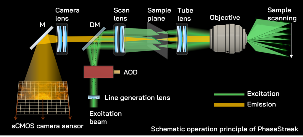
PhaseStreak scans a focused excitation line across the sample using an acousto-optic deflector synchronised with the rolling shutter of a high QE sCMOS camera. The active sensor rows form an electronically adjustable confocal detection slit that suppresses out-of-focus fluorescence and enables optimizing the balance between background rejection and signal collection for each sample.

AOD-BASED SCANNER

Fast, precise and inertia-free control of excitation laser beam.

PARALLEL LINE ACQUISITION

An entire line of pixels is acquired simultaneously, enabling confocal imaging at demonstrated speeds of up to 230 fps.

LOW-MAINTENANCE ARCHITECTURE

With no mechanical scanners or physical pinholes, PhaseStreak minimizes alignment drift, mechanical wear and avoids dust-related pinhole contamination.

03/ DEEP-SAMPLE IMAGING

See deeper
Preserve contrast

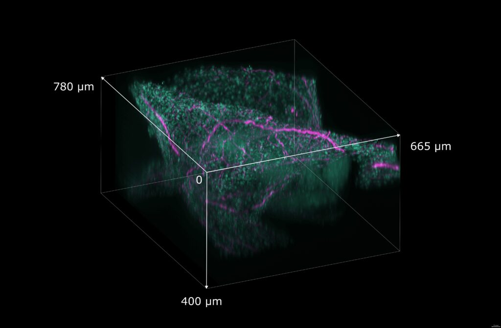
As imaging depth increases, scattering and out-of-focus fluorescence progressively reduce contrast. PhaseStreak combines selective confocal detection with efficient signal collection to preserve image clarity throughout thick samples.

3D volumetric view of a cleared mouse liver tissue down to 400 µm. Sample: 550 µm cleared mouse liver section; SYTOX Orange/nuclei (green), Alexa Fluor 647-TUJ1/nervous system (magenta).

04 / GENTLE LIVE-CELL IMAGING

Taking care of your cells

Following biological dynamics over time requires a careful balance between image quality and sample exposure. PhaseStreak gives you direct control over where excitation is delivered and how fluorescence is collected, helping you adapt that balance to each experiment.

01

>300 SIGNAL-TO-BACKGROUND RATIO

Achieve high image contrast with lower laser power.

02

DISTRIBUTED LINE EXCITATION

Distributed line illumination and efficient signal acquisition minimise the illumination dose required for high-contrast imaging.

03

ROI ILLUMINATION

Illuminate only the area you need while increasing acquisition speeds up to the kHz range.

04

TUNABLE SIGNAL COLLECTION

Adapt optical sectioning and signal collection to each sample.

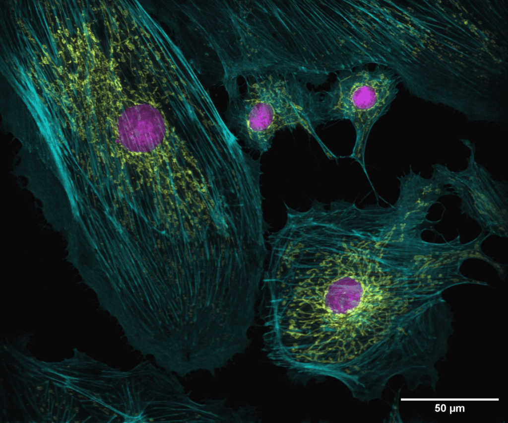
Multicolor confocal image of BPAE cells acquired with PhaseStreak. Nuclei are labelled with DAPI (magenta), F-actin with Alexa Fluor 488 phalloidin (cyan) and mitochondria with MitoTracker Red CMXRos (yellow). Sample: Thermo Fisher FluoCells Prepared Slide #1, F36924. Objective: Plan Apo 60x WI NA1.2

05 / SHARPEYE SOFTWARE

Control Automate Acquire
All from one interface

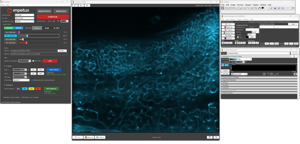
SharpEye brings all PhaseStreak controls and automated acquisition workflows into a single, self-contained interface, combining streamlined operation with the flexibility of the Micro-Manager ecosystem.

INSTANT MODE
SWITCHING

Switch between confocal, widefield and brightfield imaging in one click.

ELECTRONIC SECTIONING CONTROL

Adjust the confocal detection aperture and tune signal collection directly from the interface.

FOUR-CHANNEL
ACQUISITION

Control laser power independently at 405, 473, 561 and 639 nm and configure sequential multichannel acquisition.

AUTOMATED
ACQUISITION

Configure and run multichannel Z-stacks, time-lapse recordings and acquisition sequences from the same workspace.

Compatible with established Fiji/ImageJ visualisation and analysis workflows.

06 / SYSTEM INTEGRATION

Add confocal imaging to your setup

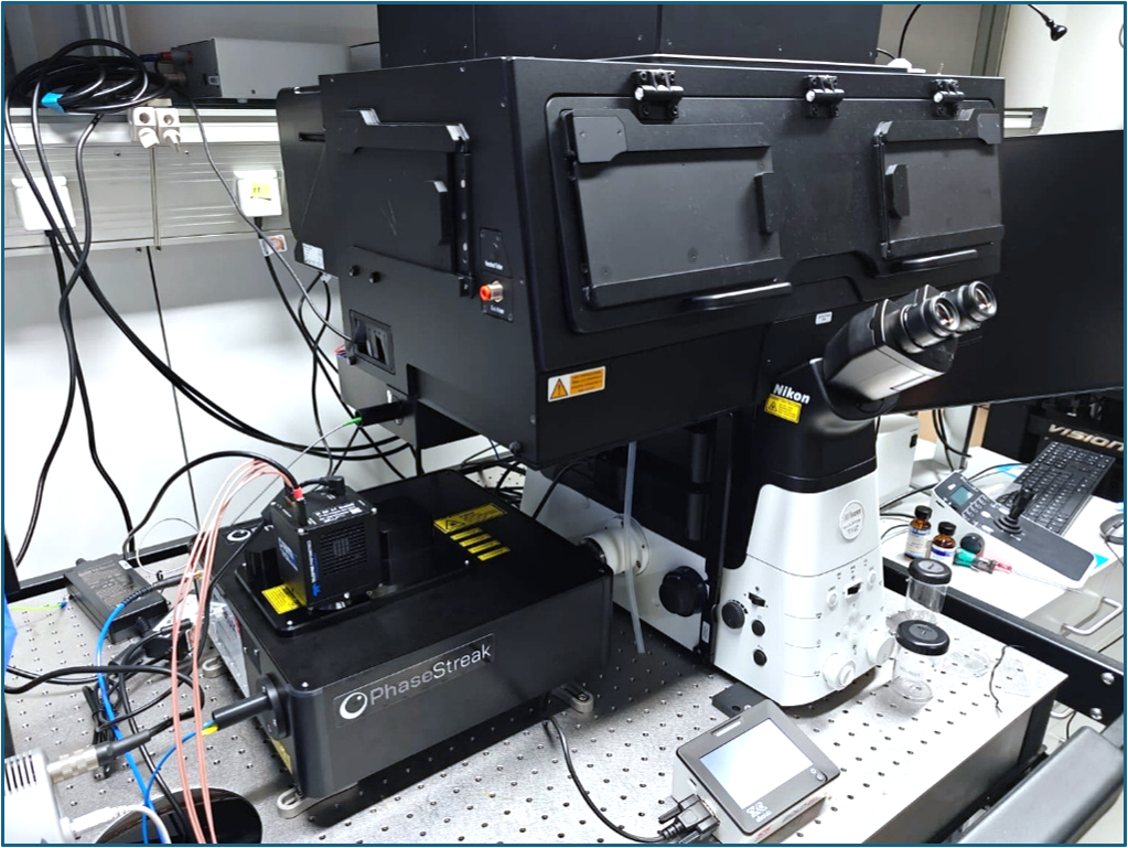
PhaseStreak integrates via the camera port of compatible inverted microscopes and can also be combined with SENSOCELL+ for correlated force and fluorescence measurements.

07 / KEY SPECIFICATIONS

Phasestreak at a glance

230 FPS
Confocal imaging speed*
23.1 mm FOV
Maximum tested camera field of view**
300+ µm
Demonstrated imaging depth
>300
Signal-to-background ratio
Solid-state architecture
No mechanical scanners or physical pinholes
Instant mode switch
Confocal, widefield and brightfield
4 excitation lines
405, 473, 561 and 639 nm
Electronic control
Tunable confocal sectioning and ROI illumination with kHz-range acquisition

* Acquisition speed is camera-limited. Demonstrated confocal imaging at 230 fps using a Tucsen Aries 6506 camera in Speed mode over 2400 × 2048 pixels, corresponding to 20.5 mm diagonal field of view.

** Demonstrated with a Hamamatsu ORCA-Fire camera over its full 4432 × 2368 pixel sensor area.
08 / SEE IT IN ACTION

Experience a new approach to confocal imaging

Discover how PhaseStreak’s solid-state architecture brings greater speed, flexibility and control to your imaging workflows.

Fig. 1

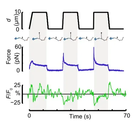
Time dependence of trap position (top), force signal (middle) and Ca2+ fluorescence signal (bottom) recorded during a membrane tether pulling experiment. The force and Ca2+ ion channel activity signals exhibit negative correlation.

Fig. 2

Force and displacement data for two beads stimulated at frequencies of 4 Hz and 0.25 Hz. The progressive reduction of the beads displacement is in concomitancy with an increase of the applied force and loading rate.

Fig. 1

Schematic representation of the optical tweezers experiment. A fibronectin-coated bead is trapped, brought into contact with the cell membrane and stimulated with oscillations at different frequencies.

Fig.2

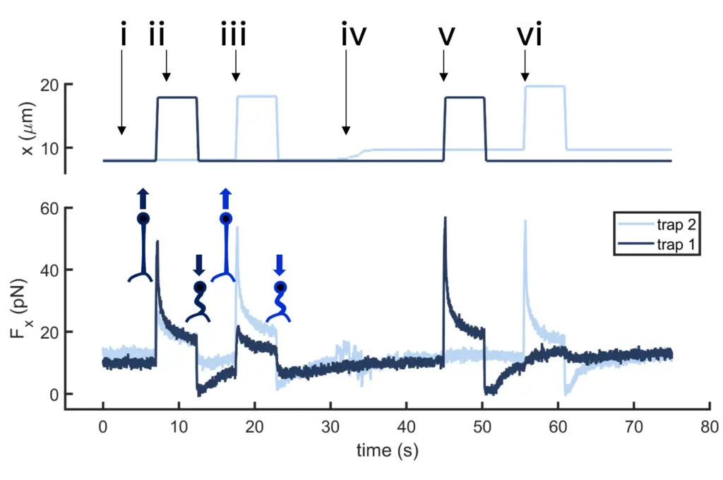
Time dependence of trap 1 and trap 2 position (top) and force (bottom) data along the different steps of the described dual tether pulling experiment.

Fig.1

Confocal video of the described dual tether pulling assay performed on a neuron axon. Courtesy of M. Krieg lab (ICFO).

Fig. 1

Time dependence of trap position (top), force signal (middle) and Ca2+ fluorescence signal (bottom) recorded during a membrane tether pulling experiment. The force and Ca2+ ion channel activity signals exhibit negative correlation.

Video 1

Confocal video showing a DVA neuron Ca2+ ion channel activity in a dynamic optical trapping assay. The pulling rate applied to the membrane tether is progressively increased. Scale bar = 5µm. Acquired at 10Hz.